
- د.حمدينو 2025
- د.حمدينو 2025
- dr.M Hussein 2026Pericardium, internal & external features
- dr.M Hussein 2026Pericardium, internal & external features
- د.أيمن خنفور
- dr.simplifiedدكتور زميلنا بيشرح على كليتنا
- dr.simplified
- ملخص 2025
2024
- Mitral valve disease cause dysphagia
2025
internal features of Rt atrium "written"
Venae cordis minimi open in all heart chambers.
Enumerate papillary muscles. Left ventricle contain two (anterior and posterior) while right ventricle three (+ septal).
2026
I. The Pericardium: Structure and Function
- The pericardium is a fibroserous sac surrounding the heart.
- Composed of two main parts:
- Fibrous pericardium (outer layer).
- Serous pericardium (inner layer).
- The serous pericardium has:
- Parietal layer: adheres to fibrous pericardium.
- Visceral layer: adheres to heart surface = Epicardium and coverd heart.
- The heart enters the serous pericardium from above and behind → visceral layer sticks to the heart, parietal layer to the fibrous layer.
Pericardial Orientation and Attachments
- Heart: pyramidal, apex forward and left; base posterior.
- Fibrous pericardium: opposite orientation, base inferior (attached to central tendon of diaphragm), apex superior (fused with great vessels).
Functions
- Fibrous pericardium:
- Prevents overdistension of the heart.
- Fixes heart in position in mediastinum.
- Provides protection against shock.
- Serous pericardium:
- Allows frictionless movement during contraction and relaxation.
Neurovascular Supply
- Blood supply:
- Outer layers (fibrous + parietal): pericardiacophrenic and muscular branches of internal thoracic artery.
- Inner layer (visceral/epicardium): coronary artery same heart.
- Nerve supply:
- Autonomic.
- Outer layers sensitive to pain (phrenic nerve C3–C5).
- Visceral layer insensitive to pain.
- Clinical: ischemia in myocardium causes angina (pain due to poor blood supply).
II. Pericardial Sinuses
- Transverse sinus
- Between great arteries (aorta, pulmonary trunk) and veins (atria).
- Lies anterior to atria, posterior to aorta and pulmonary trunk.
- Right pulmonary artery runs above it.
- Surgeon can pass finger or instrument through it.
- Oblique sinus
- On posterior surface of left atrium.
- Between right and left pulmonary veins, behind base of heart.
- Opens downward, extends upward.
- Formed during development of heart tube and serous pericardium.
- Relations: base of left atrium posteriorly, right atrium to right, left atrium to left.
III. External Features of the Heart
- General shape: conical or pyramidal.
Apex
- Directed forward, downward, left.
- At 5th left intercostal space, about 9 cm from midline.
- Formed mainly by left ventricle.
Base
- Directed posteriorly.
- Formed mainly by left atrium, partly right atrium.
- Receives four pulmonary veins.
- Contains coronary sinus draining into right atrium.
Borders of the Heart
- Right border: formed by right atrium (SVC → IVC), convex, sulcus terminalis externally.
- Inferior border: formed mainly by right ventricle, convex.
- Left border: formed mainly by left ventricle, from left auricle to apex.
- Superior border: connects right and left sides.
Surfaces
- Anterior (sternocostal): mostly right ventricle, includes anterior interventricular sulcus (LAD artery).
- Diaphragmatic (inferior): left ventricle + part of right ventricle, posterior interventricular sulcus (posterior interventricular artery + middle cardiac vein).
- Left pulmonary surface: mainly left ventricle, separated from left atrium by obtuse margin, left marginal artery runs along margin.
- Right pulmonary surface: mainly right atrium, coronary sulcus here with right coronary artery.
- Coronary sulcus: between atria and ventricles; contains left coronary artery and great cardiac vein.
IV. Internal Structure of the Right Atrium
- Has smooth posterior part (sinus venarum) and rough anterior part (pectinate muscles).
- Crista terminalis separates smooth and rough parts.
- Corresponds externally to sulcus terminalis.
- Pectinate muscles more prominent in auricle.
- Openings (5):
- SVC.
- IVC.
- Tricuspid valve (right AV orifice).
- Coronary sinus.
- Small veins (venae cordis minimae).
- Interatrial septum features:
- Fossa ovalis (depression).
- Limbus fossa ovalis (prominent margin).
V. Internal Structure of the Right Ventricle
- Smooth part (outflow): infundibulum (conus arteriosus) → pulmonary artery.
- Rough part (inflow): trabeculae carneae, papillary muscles (3: anterior, posterior, septal).
- Chordae tendineae attach papillary muscles to tricuspid valve cusps.
- Moderator band connects septum to wall, carries right bundle branch.
- Valves:
- Tricuspid valve (3 cusps).
- Pulmonary valve (3 cusps): 1 posterior, 2 anterior.
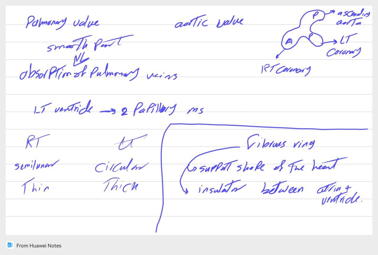
Semilunar Valves Comparison
- Pulmonary valve: 1 posterior, 2 anterior cusps.
- Aortic valve: 1 anterior, 2 posterior cusps.
- Aortic sinuses above cusps → right coronary artery from anterior sinus, left coronary from left posterior sinus.
VI. Internal Structure of the Left Atrium
- Smooth posterior part formed by absorbed pulmonary veins.
- Rough part (pectinate muscles) confined to auricle.
- Receives 4 pulmonary veins.
- Orifices: pulmonary veins + left AV (mitral) valve.
VII. Internal Structure of the Left Ventricle
- Outflow tract: smooth (aortic vestibule).
- Inflow tract: trabeculae carneae + 2 papillary muscles.
- Wall much thicker than right ventricle.
- Left ventricle pumps to systemic circulation → higher pressure.
- IV septum bulges to right → right ventricle crescent-shaped.
VIII. Cardiac Skeleton and Conduction System
Cardiac Skeleton
- Fibrous connective tissue framework supporting valves (aortic, pulmonary, tricuspid, mitral).
- Functions:
- Anchors valve cusps and myocardium.
- Acts as electrical insulator between atria and ventricles.
Conduction System
- Impulse starts at SA node → AV node → Bundle of His → ventricles.
- Fibrous skeleton prevents direct spread of impulse from atria to ventricles.
- AV node and bundle provide only conductive connection, ensuring sequential contraction (atria first, then ventricles).
2024
- Mitral valve disease cause dysphagia
2025
internal features of Rt atrium "written"
Venae cordis minimi open in all heart chambers.
Enumerate papillary muscles. Left ventricle contain two (anterior and posterior) while right ventricle three (+ septal).
2026
I. The Pericardium: Structure and Function
- The pericardium is a fibroserous sac surrounding the heart.
- Composed of two main parts:
- Fibrous pericardium (outer layer).
- Serous pericardium (inner layer).
- The serous pericardium has:
- Parietal layer: adheres to fibrous pericardium.
- Visceral layer: adheres to heart surface = Epicardium and coverd heart.
- The heart enters the serous pericardium from above and behind → visceral layer sticks to the heart, parietal layer to the fibrous layer.
Pericardial Orientation and Attachments
- Heart: pyramidal, apex forward and left; base posterior.
- Fibrous pericardium: opposite orientation, base inferior (attached to central tendon of diaphragm), apex superior (fused with great vessels).
Functions
- Fibrous pericardium:
- Prevents overdistension of the heart.
- Fixes heart in position in mediastinum.
- Provides protection against shock.
- Serous pericardium:
- Allows frictionless movement during contraction and relaxation.
Neurovascular Supply
- Blood supply:
- Outer layers (fibrous + parietal): pericardiacophrenic and muscular branches of internal thoracic artery.
- Inner layer (visceral/epicardium): coronary artery same heart.
- Nerve supply:
- Autonomic.
- Outer layers sensitive to pain (phrenic nerve C3–C5).
- Visceral layer insensitive to pain.
- Clinical: ischemia in myocardium causes angina (pain due to poor blood supply).
II. Pericardial Sinuses
- Transverse sinus
- Between great arteries (aorta, pulmonary trunk) and veins (atria).
- Lies anterior to atria, posterior to aorta and pulmonary trunk.
- Right pulmonary artery runs above it.
- Surgeon can pass finger or instrument through it.
- Oblique sinus
- On posterior surface of left atrium.
- Between right and left pulmonary veins, behind base of heart.
- Opens downward, extends upward.
- Formed during development of heart tube and serous pericardium.
- Relations: base of left atrium posteriorly, right atrium to right, left atrium to left.
III. External Features of the Heart
- General shape: conical or pyramidal.
Apex
- Directed forward, downward, left.
- At 5th left intercostal space, about 9 cm from midline.
- Formed mainly by left ventricle.
Base
- Directed posteriorly.
- Formed mainly by left atrium, partly right atrium.
- Receives four pulmonary veins.
- Contains coronary sinus draining into right atrium.
Borders of the Heart
- Right border: formed by right atrium (SVC → IVC), convex, sulcus terminalis externally.
- Inferior border: formed mainly by right ventricle, convex.
- Left border: formed mainly by left ventricle, from left auricle to apex.
- Superior border: connects right and left sides.
Surfaces
- Anterior (sternocostal): mostly right ventricle, includes anterior interventricular sulcus (LAD artery).
- Diaphragmatic (inferior): left ventricle + part of right ventricle, posterior interventricular sulcus (posterior interventricular artery + middle cardiac vein).
- Left pulmonary surface: mainly left ventricle, separated from left atrium by obtuse margin, left marginal artery runs along margin.
- Right pulmonary surface: mainly right atrium, coronary sulcus here with right coronary artery.
- Coronary sulcus: between atria and ventricles; contains left coronary artery and great cardiac vein.
IV. Internal Structure of the Right Atrium
- Has smooth posterior part (sinus venarum) and rough anterior part (pectinate muscles).
- Crista terminalis separates smooth and rough parts.
- Corresponds externally to sulcus terminalis.
- Pectinate muscles more prominent in auricle.
- Openings (5):
- SVC.
- IVC.
- Tricuspid valve (right AV orifice).
- Coronary sinus.
- Small veins (venae cordis minimae).
- Interatrial septum features:
- Fossa ovalis (depression).
- Limbus fossa ovalis (prominent margin).
V. Internal Structure of the Right Ventricle
- Smooth part (outflow): infundibulum (conus arteriosus) → pulmonary artery.
- Rough part (inflow): trabeculae carneae, papillary muscles (3: anterior, posterior, septal).
- Chordae tendineae attach papillary muscles to tricuspid valve cusps.
- Moderator band connects septum to wall, carries right bundle branch.
- Valves:
- Tricuspid valve (3 cusps).
- Pulmonary valve (3 cusps): 1 posterior, 2 anterior.
Semilunar Valves Comparison
- Pulmonary valve: 1 posterior, 2 anterior cusps.
- Aortic valve: 1 anterior, 2 posterior cusps.
- Aortic sinuses above cusps → right coronary artery from anterior sinus, left coronary from left posterior sinus.
VI. Internal Structure of the Left Atrium
- Smooth posterior part formed by absorbed pulmonary veins.
- Rough part (pectinate muscles) confined to auricle.
- Receives 4 pulmonary veins.
- Orifices: pulmonary veins + left AV (mitral) valve.
VII. Internal Structure of the Left Ventricle
- Outflow tract: smooth (aortic vestibule).
- Inflow tract: trabeculae carneae + 2 papillary muscles.
- Wall much thicker than right ventricle.
- Left ventricle pumps to systemic circulation → higher pressure.
- IV septum bulges to right → right ventricle crescent-shaped.
VIII. Cardiac Skeleton and Conduction System
Cardiac Skeleton
- Fibrous connective tissue framework supporting valves (aortic, pulmonary, tricuspid, mitral).
- Functions:
- Anchors valve cusps and myocardium.
- Acts as electrical insulator between atria and ventricles.
Conduction System
- Impulse starts at SA node → AV node → Bundle of His → ventricles.
- Fibrous skeleton prevents direct spread of impulse from atria to ventricles.
- AV node and bundle provide only conductive connection, ensuring sequential contraction (atria first, then ventricles).