
- د. حمدينو 2026
- د. حمدينو 2026
- د.ايمن خنفور
- ملخص 2026الصباغ
- ملخص 2026R 🦋
🖇 I. MUSCLES OF THE NECK
📌 A. Suprahyoid Muscles (Above the Hyoid Bone)
Function: Suspend the Hyoid bone from above, positioned between the Mandible and Hyoid bone.
Four Muscles:
- Digastric Muscle
- Named for two gaster (two heads/bellies)
- Origin: One head from Mandible, other from Mastoid Process
- Stylohyoid Muscle
- Origin: Styloid Process
- Insertion: Hyoid bone
- Geniohyoid Muscle
- Origin: Mandible (specifically Genial Tubercle)
- Insertion: Hyoid bone
- Mylohyoid Muscle
- Attachment: Entire length of Mandible to Hyoid bone
📌 B. Infrahyoid Muscles (Below the Hyoid Bone)
Function: Connect Hyoid bone downwards to Sternum, Clavicle, and Scapula, passing the Thyroid cartilage (Adam's apple).
Arrangement: Two layers
Four Muscles:
- Thyrohyoid Muscle
- Connection: Thyroid cartilage ↔ Hyoid bone
- Sternothyroid Muscle
- Connection: Thyroid cartilage ↔ Sternum
- Sternohyoid Muscle
- Connection: Hyoid bone directly ↔ Sternum
- Omohyoid Muscle
- Connection: Hyoid ↔ Scapula
- Structure: Two bellies (superior and inferior), similar to Digastric
🖇 II. GENERAL ANATOMY AND TRIANGLES OF THE NECK
📌 A. Other Muscle Groups
Prevertebral Muscles:
- Located anteriorly, related to vertebral column
- Extensive details not covered in this lecture
Lateral Muscles:
- Scalene muscles: Scalenus Anterior, Scalenus Medius, Scalenus Posterior
Sternocleidomastoid Muscle (SCM):
- Oblique muscle
- Course: Sternum and Clavicle → Mastoid process
📌 B. Visceral Spaces
Location: Between Hyoid bone and surrounding muscles
Contents:
- Fascial space containing viscera
- Thyroid gland
- Carotid sheath with its contents
🚨 C. Posterior Triangle Subdivision (Exam Point)
Key Dividing Structure:
- Inferior Belly of Omohyoid Muscle runs horizontally across Posterior Triangle
Division Result: Two smaller triangles:
- Superior Triangle = Occipital Triangle
- Inferior Triangle = Supraclavicular Triangle (located above clavicle)
Complexity Note:
- Anterior Triangle is MORE complex than Posterior Triangle
- Reason: Complexity created by Hyoid bone and related musculature
📌 D. Boundaries, Roof, and Floor of Posterior Triangle
🟠 1. The Roof
Superficial Layer:
- Skin
- Superficial Fascia beneath skin
Superficial Fascia Contents:
- Platysma muscle (muscle of facial expression)
- Cutaneous nerves
Cutaneous Nerve Origin:
- ALL cutaneous nerves in neck = branches of Cervical Plexus
Deep Fascia (Cervical Fascia): Complex structure = three tubes nested within each other:
- Investing Layer (outermost)
- Envelops entire neck
- Pretracheal Fascia (middle tube)
- Contains viscera (e.g., Thyroid gland)
- Prevertebral Fascia (innermost tube)
- Surrounds vertebral column and attached muscles
🟠 2. The Floor
Composition: Four muscle layers resting laterally on vertebral column
Muscles (superficial to deep):
- Semispinalis Capitis
- Splenius Capitis
- Levator Scapulae
- Scalenus Medius
- Note: Scalenus Posterior may be present (varies with dissection)
🚨 Fascial Covering: Prevertebral Fascia covers muscular floor (Exam Point)
📌 E. Contents of Posterior Triangle (Neurovascular)
🟣 Veins:
- External Jugular Vein (EJV)
🟣 Arteries:
Subclavian Artery:
- Third Part of Subclavian Artery present in triangle
- Division mechanism: Scalenus Anterior Muscle passes in front of Subclavian Artery
- Creates three parts:
- 1st part: medial to Scalenus Anterior
- 2nd part: posterior to Scalenus Anterior
- 3rd part: lateral to Scalenus Anterior
Thyrocervical Trunk (TCT):
- Branch of first part of Subclavian Artery
- Branches:
- Superior branch: Inferior Thyroid Artery
- Two anterior branches:
- Transverse Cervical Artery
- Suprascapular Artery
🖇 III. SPINAL NERVES AND PLEXUSES (CRUCIAL CONCEPT)
📌 A. Spinal Nerve Organization
Total: 31 pairs of spinal nerves
Immediate Division upon exiting spinal column:
1. Dorsal Ramus:
- Immediately enters Epaxial Muscles (Intrinsic back muscles)
- Straightforward course
- Do NOT form plexuses
2. Ventral Ramus (Larger):
- Supplies all other muscles (Hypaxial Muscles)
- Includes: trunk muscles, limb muscles, muscles lying directly on vertebrae (even if posterior)
🚨 B. Mechanism and Purpose of Plexuses (Exam Point)
Definition:
- Nerve networks formed when multiple nerve roots merge, branch, merge again, branch again
Purpose (Triple Redundancy):
- Single muscle supplied by multiple nerve roots
- Single nerve root supplies multiple muscles
- If one innervation route damaged → guaranteed supply maintained via another route
📌 C. Spinal Cord Enlargements
Reason: Segments where ventral rami contribute to limb plexuses are enlarged due to increased neuronal volume needed to supply limbs (structures "added on trunk")
Two Enlargements:
- Cervical Enlargement (C5–T1)
- For Brachial Plexus (Upper Limb)
- Lumbosacral Enlargement (L1–S3)
- For Lumbosacral Plexus (Lower Limb)
📌 D. Rules for Plexus Formation
Question: Do ALL Ventral Rami form plexuses? Answer: NO
🚨 Thoracic Exception (T1–T12):
- Thoracic ventral rami do NOT form plexuses
- Reason: Ribs act as "concrete walls" preventing nerve communication/mixing
- Result: Nerves remain isolated, travel individually in intercostal spaces
Plexuses Above and Below Thorax:
- Cervical Plexus (C1–C4)
- Supplies: neck, part of face, Phrenic nerve (diaphragm), deep neck muscles
- Brachial Plexus (C5–T1)
- Supplies: upper limb
- Lumbar Plexus (L1–L4)
- Supplies: pelvis, perineum, lower limb
- Sacral Plexus (L4, L5, S1, S2, S3, part of S4)
- Supplies: pelvis, perineum, lower limb
- Coccygeal Plexus (Part of S4, S5, Coccygeal Nerve)
- Supplies: perineum
🖇 IV. CRANIAL NERVE FIBERS CLASSIFICATION
🚨 CRITICAL EXAM POINT: This section MORE IMPORTANT than entire lecture material
📌 A. Classification Table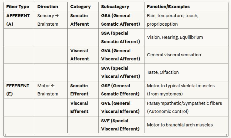 🟠 B. Special Visceral Efferent (SVE) System - Detailed
Distinct Class: Separate from other motor systems
Target Muscles:
- Muscles derived from Branchial Arches (Pharyngeal Arches)
- Includes: Pharynx muscles, Larynx muscles, Mastication muscles
Functional Nature (Hybrid):
- Part somatic-like: Can be initiated consciously
- Part visceral-like: Involuntary
- Example: Swallowing
- First stage: voluntary (somatic-like)
- Subsequent stages: involuntary (visceral-like)
Motor Nuclei:
- Nucleus Ambiguus (medulla oblongata)
- Responsible for many SVE fibers
- Supplies: pharynx/larynx muscles
- Motor Nucleus of Trigeminal Nerve
- Supplies: mastication muscles (from 1st branchial arch)
🖇 V. STERNOCLEIDOMASTOID MUSCLE (SCM) - DETAILED STUDY
📌 A. Anatomical Details
Origin (Two Heads):
- Sternal head
- Clavicular head
Insertion:
- Outer surface of Mastoid Process
🚨 Nerve Supply: Spinal Accessory Nerve (CN XI) (Exam Point)
🚨 Unique Feature: SCM and Trapezius = ONLY two skeletal muscles in neck supplied by cranial nerve (Exam Point)
📌 B. Spinal Accessory Nerve (CN XI) Composition
Two Components:
1. Cranial Root:
- Origin: Nucleus Ambiguus
2. Spinal Root (Spinal Accessory N.):
- Origin: Central nucleus in spinal cord (C1–C5 segments)
Course:
- Two roots travel together without mixing
- Exit via Jugular Foramen
- Then separate:
Spinal Root Distribution (CN XI spinal portion):
- Supplies ONLY:
- Sternocleidomastoid muscle
- Trapezius muscle
Cranial Root Distribution (CN XI cranial portion):
- Joins Vagus nerve (CN X)
- Supplies: muscles from branchial arches (Pharynx and Larynx)
- Name "Accessory" = because it adds these fibers to Vagus
📌 C. Functional Significance and Coordination
Primary Function: Largely involved in reflex action
- Justifies cranial nerve supply
Example: Visual-Audio Spinal Reflex
- Coordination needed between:
- Eyes
- Facial muscles
- Neck muscles
- Situation: Reacting to stimuli or tracking objects (e.g., watching scenery from moving train)
Coordinating Structure: Medial Longitudinal Fasciculus (MLF)
- Functions as "electrical cable"
- Connects nuclei from Midbrain → Spinal Cord:
- Oculomotor nucleus
- Abducens nucleus
- Trochlear nucleus
- Nucleus Ambiguus
- Facial nucleus
- Result: Coordinated action between:
- Extraocular muscles
- Facial muscles
- SCM (via Spinal Accessory Nerve)
🚨 D. Actions of SCM (Exam Points)
Bilateral Contraction (Both SCMs):
- Action: Extension of Head
🚨 Unilateral Contraction (One SCM): (Exam Point)
- Lateral Flexion (tilting ear toward shoulder on same side)
- Face rotates to OPPOSITE side (outward rotation)
🚨 SCM and Contralateral Movement Principle: (Exam Point)
- SCM works contralaterally to trunk muscles
- Brain-body relationship:
- Right brain controls left body
- Left arm active = Right brain working
- To look toward Left arm = Right SCM must contract
🖇 I. MUSCLES OF THE NECK
📌 A. Suprahyoid Muscles (Above the Hyoid Bone)
Function: Suspend the Hyoid bone from above, positioned between the Mandible and Hyoid bone.
Four Muscles:
- Digastric Muscle
- Named for two gaster (two heads/bellies)
- Origin: One head from Mandible, other from Mastoid Process
- Stylohyoid Muscle
- Origin: Styloid Process
- Insertion: Hyoid bone
- Geniohyoid Muscle
- Origin: Mandible (specifically Genial Tubercle)
- Insertion: Hyoid bone
- Mylohyoid Muscle
- Attachment: Entire length of Mandible to Hyoid bone
📌 B. Infrahyoid Muscles (Below the Hyoid Bone)
Function: Connect Hyoid bone downwards to Sternum, Clavicle, and Scapula, passing the Thyroid cartilage (Adam's apple).
Arrangement: Two layers
Four Muscles:
- Thyrohyoid Muscle
- Connection: Thyroid cartilage ↔ Hyoid bone
- Sternothyroid Muscle
- Connection: Thyroid cartilage ↔ Sternum
- Sternohyoid Muscle
- Connection: Hyoid bone directly ↔ Sternum
- Omohyoid Muscle
- Connection: Hyoid ↔ Scapula
- Structure: Two bellies (superior and inferior), similar to Digastric
🖇 II. GENERAL ANATOMY AND TRIANGLES OF THE NECK
📌 A. Other Muscle Groups
Prevertebral Muscles:
- Located anteriorly, related to vertebral column
- Extensive details not covered in this lecture
Lateral Muscles:
- Scalene muscles: Scalenus Anterior, Scalenus Medius, Scalenus Posterior
Sternocleidomastoid Muscle (SCM):
- Oblique muscle
- Course: Sternum and Clavicle → Mastoid process
📌 B. Visceral Spaces
Location: Between Hyoid bone and surrounding muscles
Contents:
- Fascial space containing viscera
- Thyroid gland
- Carotid sheath with its contents
🚨 C. Posterior Triangle Subdivision (Exam Point)
Key Dividing Structure:
- Inferior Belly of Omohyoid Muscle runs horizontally across Posterior Triangle
Division Result: Two smaller triangles:
- Superior Triangle = Occipital Triangle
- Inferior Triangle = Supraclavicular Triangle (located above clavicle)
Complexity Note:
- Anterior Triangle is MORE complex than Posterior Triangle
- Reason: Complexity created by Hyoid bone and related musculature
📌 D. Boundaries, Roof, and Floor of Posterior Triangle
🟠 1. The Roof
Superficial Layer:
- Skin
- Superficial Fascia beneath skin
Superficial Fascia Contents:
- Platysma muscle (muscle of facial expression)
- Cutaneous nerves
Cutaneous Nerve Origin:
- ALL cutaneous nerves in neck = branches of Cervical Plexus
Deep Fascia (Cervical Fascia): Complex structure = three tubes nested within each other:
- Investing Layer (outermost)
- Envelops entire neck
- Pretracheal Fascia (middle tube)
- Contains viscera (e.g., Thyroid gland)
- Prevertebral Fascia (innermost tube)
- Surrounds vertebral column and attached muscles
🟠 2. The Floor
Composition: Four muscle layers resting laterally on vertebral column
Muscles (superficial to deep):
- Semispinalis Capitis
- Splenius Capitis
- Levator Scapulae
- Scalenus Medius
- Note: Scalenus Posterior may be present (varies with dissection)
🚨 Fascial Covering: Prevertebral Fascia covers muscular floor (Exam Point)
📌 E. Contents of Posterior Triangle (Neurovascular)
🟣 Veins:
- External Jugular Vein (EJV)
🟣 Arteries:
Subclavian Artery:
- Third Part of Subclavian Artery present in triangle
- Division mechanism: Scalenus Anterior Muscle passes in front of Subclavian Artery
- Creates three parts:
- 1st part: medial to Scalenus Anterior
- 2nd part: posterior to Scalenus Anterior
- 3rd part: lateral to Scalenus Anterior
Thyrocervical Trunk (TCT):
- Branch of first part of Subclavian Artery
- Branches:
- Superior branch: Inferior Thyroid Artery
- Two anterior branches:
- Transverse Cervical Artery
- Suprascapular Artery
🖇 III. SPINAL NERVES AND PLEXUSES (CRUCIAL CONCEPT)
📌 A. Spinal Nerve Organization
Total: 31 pairs of spinal nerves
Immediate Division upon exiting spinal column:
1. Dorsal Ramus:
- Immediately enters Epaxial Muscles (Intrinsic back muscles)
- Straightforward course
- Do NOT form plexuses
2. Ventral Ramus (Larger):
- Supplies all other muscles (Hypaxial Muscles)
- Includes: trunk muscles, limb muscles, muscles lying directly on vertebrae (even if posterior)
🚨 B. Mechanism and Purpose of Plexuses (Exam Point)
Definition:
- Nerve networks formed when multiple nerve roots merge, branch, merge again, branch again
Purpose (Triple Redundancy):
- Single muscle supplied by multiple nerve roots
- Single nerve root supplies multiple muscles
- If one innervation route damaged → guaranteed supply maintained via another route
📌 C. Spinal Cord Enlargements
Reason: Segments where ventral rami contribute to limb plexuses are enlarged due to increased neuronal volume needed to supply limbs (structures "added on trunk")
Two Enlargements:
- Cervical Enlargement (C5–T1)
- For Brachial Plexus (Upper Limb)
- Lumbosacral Enlargement (L1–S3)
- For Lumbosacral Plexus (Lower Limb)
📌 D. Rules for Plexus Formation
Question: Do ALL Ventral Rami form plexuses? Answer: NO
🚨 Thoracic Exception (T1–T12):
- Thoracic ventral rami do NOT form plexuses
- Reason: Ribs act as "concrete walls" preventing nerve communication/mixing
- Result: Nerves remain isolated, travel individually in intercostal spaces
Plexuses Above and Below Thorax:
- Cervical Plexus (C1–C4)
- Supplies: neck, part of face, Phrenic nerve (diaphragm), deep neck muscles
- Brachial Plexus (C5–T1)
- Supplies: upper limb
- Lumbar Plexus (L1–L4)
- Supplies: pelvis, perineum, lower limb
- Sacral Plexus (L4, L5, S1, S2, S3, part of S4)
- Supplies: pelvis, perineum, lower limb
- Coccygeal Plexus (Part of S4, S5, Coccygeal Nerve)
- Supplies: perineum
🖇 IV. CRANIAL NERVE FIBERS CLASSIFICATION
🚨 CRITICAL EXAM POINT: This section MORE IMPORTANT than entire lecture material
📌 A. Classification Table🟠 B. Special Visceral Efferent (SVE) System - Detailed
Distinct Class: Separate from other motor systems
Target Muscles:
- Muscles derived from Branchial Arches (Pharyngeal Arches)
- Includes: Pharynx muscles, Larynx muscles, Mastication muscles
Functional Nature (Hybrid):
- Part somatic-like: Can be initiated consciously
- Part visceral-like: Involuntary
- Example: Swallowing
- First stage: voluntary (somatic-like)
- Subsequent stages: involuntary (visceral-like)
Motor Nuclei:
- Nucleus Ambiguus (medulla oblongata)
- Responsible for many SVE fibers
- Supplies: pharynx/larynx muscles
- Motor Nucleus of Trigeminal Nerve
- Supplies: mastication muscles (from 1st branchial arch)
🖇 V. STERNOCLEIDOMASTOID MUSCLE (SCM) - DETAILED STUDY
📌 A. Anatomical Details
Origin (Two Heads):
- Sternal head
- Clavicular head
Insertion:
- Outer surface of Mastoid Process
🚨 Nerve Supply: Spinal Accessory Nerve (CN XI) (Exam Point)
🚨 Unique Feature: SCM and Trapezius = ONLY two skeletal muscles in neck supplied by cranial nerve (Exam Point)
📌 B. Spinal Accessory Nerve (CN XI) Composition
Two Components:
1. Cranial Root:
- Origin: Nucleus Ambiguus
2. Spinal Root (Spinal Accessory N.):
- Origin: Central nucleus in spinal cord (C1–C5 segments)
Course:
- Two roots travel together without mixing
- Exit via Jugular Foramen
- Then separate:
Spinal Root Distribution (CN XI spinal portion):
- Supplies ONLY:
- Sternocleidomastoid muscle
- Trapezius muscle
Cranial Root Distribution (CN XI cranial portion):
- Joins Vagus nerve (CN X)
- Supplies: muscles from branchial arches (Pharynx and Larynx)
- Name "Accessory" = because it adds these fibers to Vagus
📌 C. Functional Significance and Coordination
Primary Function: Largely involved in reflex action
- Justifies cranial nerve supply
Example: Visual-Audio Spinal Reflex
- Coordination needed between:
- Eyes
- Facial muscles
- Neck muscles
- Situation: Reacting to stimuli or tracking objects (e.g., watching scenery from moving train)
Coordinating Structure: Medial Longitudinal Fasciculus (MLF)
- Functions as "electrical cable"
- Connects nuclei from Midbrain → Spinal Cord:
- Oculomotor nucleus
- Abducens nucleus
- Trochlear nucleus
- Nucleus Ambiguus
- Facial nucleus
- Result: Coordinated action between:
- Extraocular muscles
- Facial muscles
- SCM (via Spinal Accessory Nerve)
🚨 D. Actions of SCM (Exam Points)
Bilateral Contraction (Both SCMs):
- Action: Extension of Head
🚨 Unilateral Contraction (One SCM): (Exam Point)
- Lateral Flexion (tilting ear toward shoulder on same side)
- Face rotates to OPPOSITE side (outward rotation)
🚨 SCM and Contralateral Movement Principle: (Exam Point)
- SCM works contralaterally to trunk muscles
- Brain-body relationship:
- Right brain controls left body
- Left arm active = Right brain working
- To look toward Left arm = Right SCM must contract